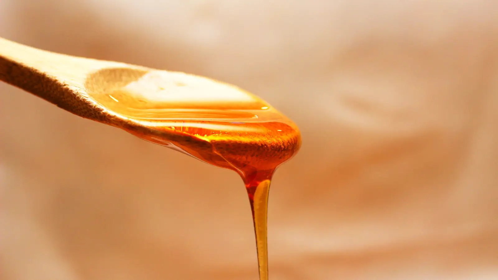
The hydrogen peroxide secret behind honey wound healing is the enzyme glucose oxidase—a bee‑deposited catalyst that, upon dilution with wound fluid, generates a slow, low‑level release of hydrogen peroxide, delivering a targeted antimicrobial clean without the tissue damage of bottled antiseptics.
Honey’s wound use predates recorded medicine, but the peroxide mechanism is neither ancient folk wisdom nor a marketing invention. It is a chemical pathway with a clear on‑off switch. Glucose oxidase enters the honey during nectar processing; in the jar, honey’s water activity hovers around 0.6, far too low for the enzyme to function. Apply that same honey to a weeping wound, and the influx of serum or interstitial fluid pushes the water activity above roughly 0.9, freeing the enzyme to bind glucose and oxygen. The reaction yields gluconic acid and hydrogen peroxide in a ratio that self‑limits. More dilution means more activation until the wound dries; less exudate slows the system down. This feedback loop is the real secret—not peroxide per se, but a rate‑controlled release that never spikes high enough to kill fibroblasts.
The enzymatic logic matters because it redefines what a wound dressing is supposed to do. Rather than flooding the injury with a fixed dose of a reactive chemical, the honey system continuously titrates the antimicrobial output against the wound’s moisture status. That makes it uniquely suited to chronic wounds stuck in the inflammatory phase, where biofilm and necrotic debris persist despite repeated antiseptic applications. Glucose oxidase doesn’t just kill bacteria; it disrupts the extracellular matrix they hide in and signals a shift in the wound’s biochemistry toward repair.
The Chemistry That Converts a Wound’s Own Moisture Into a Cleanser
Water Activity, Not Just Dilution, Dictates Enzyme Activation
Glucose oxidase is an oxidoreductase requiring both glucose and molecular oxygen. In undiluted honey, the sugar concentration is so extreme—often above 80% w/w—that water molecules are effectively sequestered in hydration shells around the sugars. The enzyme’s active site cannot access free water to catalyze the reaction. When wound exudate penetrates the honey, it raises the water activity above the critical threshold of about 0.9. At that point, the reaction becomes thermodynamically favorable and proceeds at a pace set by local glucose and oxygen diffusion. The resulting peroxide appears in the low millimolar range, a concentration that bacterial biofilms cannot easily neutralize.
Why Sustained Low‑Dose Peroxide Outperforms a Transient Bolus
A single dose of 3% hydrogen peroxide oxidizes indiscriminately, damaging not only bacterial cell walls but also the migrating keratinocytes and endothelial cells essential for closure. The honey‑driven system releases peroxide in the order of 0.1–2 mmol L⁻¹ over a period of hours, well within the hormetic window where stress responses in human cells are triggered without crossing into apoptosis. For biofilm‑dwelling bacteria, this continuous low‑grade oxidative stress overwhelms their antioxidant defenses over time while leaving the host tissue’s reparative machinery intact. The kinetics are exactly the opposite of a chemical soak.
Targeting the Biofilm Matrix Without Sharps or Heavy Enzymatic Debriders
Biofilms consist of polysaccharides, proteins, and extracellular DNA. Hydrogen peroxide at sustained low levels oxidizes the side chains of the polysaccharides, leading to chain scission and matrix depolymerization. It also interferes with quorum‑sensing autoinducers by oxidizing their thiolactone rings. The net effect is a gentle, autolytic‑style debridement that lifts the biofilm layer away from the wound bed, exposing bacteria to phagocytes and making them vulnerable to any systemic antibiotics the patient might be receiving.
Clinical Situations Where the Peroxide System Is Decisive
Infected Chronic Wounds Exhibiting Persistent Biofilm
Venous leg ulcers, pressure injuries, and diabetic foot lesions that fail to shrink despite offloading and standard dressings often harbor polymicrobial biofilms. In these wounds, the hydrogen peroxide system can be the difference between a stalled wound and one that begins to granulate. Because the enzyme activation tracks exudate production, the peroxide dose is automatically higher when the wound is most wet and infected, then tapers as healing reduces the fluid output. No external dosage adjustment is required.
Burns Where Chemical Debridement Risk Must Stay Low
Partial‑thickness burns heal by re‑epithelialization from remaining dermal appendages. Any debridement that goes too deep destroys those appendages and converts the injury to a full‑thickness defect. The glucose oxidase system offers a debridement method that stops when the eschar loosens and the exudate drops, protecting the delicate regenerating tissue below.
Contexts Where the Glucose Oxidase System Contributes Almost Nothing
Dry, Epithelializing Wounds Without Exudate
Once a wound is fully epithelialized or scabbed, there is no free water to activate the enzyme. Honey applied at this stage acts solely as an occlusive barrier, not an antimicrobial. The peroxide pathway is inert.
Honeys Where Methylglyoxal Shuts Down the Peroxide Pathway
High‑MGO manuka honeys inhibit glucose oxidase. The methylglyoxal reacts with amino acid residues in the enzyme’s active site, rendering it incapable of producing peroxide. In these products, the wound‑cleaning effect comes from a pH‑dependent and osmotic mechanism, not from enzymatic peroxide. If sustained, dilution‑triggered peroxide release is the clinical goal, manuka is effectively the wrong tool.
Concepts That Frame This Enzyme‑Driven Approach
- Glucose oxidase – A flavoprotein secreted by bee hypopharyngeal glands; its catalytic efficiency determines the peroxide‑generating capacity of any given honey.
- Medical‑grade honey – Sterilized by gamma irradiation to eliminate spores while preserving enzyme activity, allowing reliable peroxide release in clinical settings.
- Autolytic debridement – A natural process where endogenous enzymes and moisture soften and remove dead tissue; honey‑derived peroxide amplifies this without breaking the skin’s own enzyme balance.
- Quorum sensing – Bacterial communication system that coordinates biofilm formation; low‑level peroxide disrupts the signaling molecules, dismantling the cooperative defense.
Frequently Asked Questions
Does every honey contain active glucose oxidase?
No. The enzyme’s presence depends on bee species, floral source, processing, and storage temperature. Heating honey above 40°C denatures glucose oxidase, and some nectar sources yield honey naturally low in the enzyme. Only raw, unheated honeys or properly processed medical‑grade honeys retain viable enzyme activity.
If honey produces peroxide, why doesn’t it bleach skin or clothing?
The concentration is too low—rarely exceeding 2 mmol L⁻¹. Significant bleaching requires peroxide concentrations orders of magnitude higher. The same low level explains why wound pain is minimal compared to bottled peroxide.
Can I activate glucose oxidase by mixing honey with water before applying it to a wound?
This is not recommended. Pre‑diluting honey gives the enzyme substrate water, but it also triggers a rapid burst of peroxide in the container, not on the wound. The peroxide will dissipate before application, and the remaining honey may have lost most of its antimicrobial activity. Let the wound exudate activate the enzyme in situ.
Is hydrogen peroxide from honey effective against multidrug‑resistant bacteria like MRSA?
Yes, in laboratory studies honeys with high glucose oxidase activity have shown efficacy against MRSA and other resistant strains because the oxidative mechanism is nonspecific and attacks multiple bacterial targets simultaneously. However, the effect depends entirely on adequate enzyme activity and sustained contact—it is not a rapid sterilant.
Where the Next Question Leads
The natural next inquiry concerns selecting a honey product whose glucose oxidase activity is verifiable and stable under wound‑care conditions. That moves into the territory of honey testing methods—catalase interference, peroxide accumulation curves, and international standards for medical‑grade honey, which collectively answer whether a given jar will actually deliver the slow‑release peroxide logic described here.